Tips on how to manage acute ankle, knee and shoulder injuries
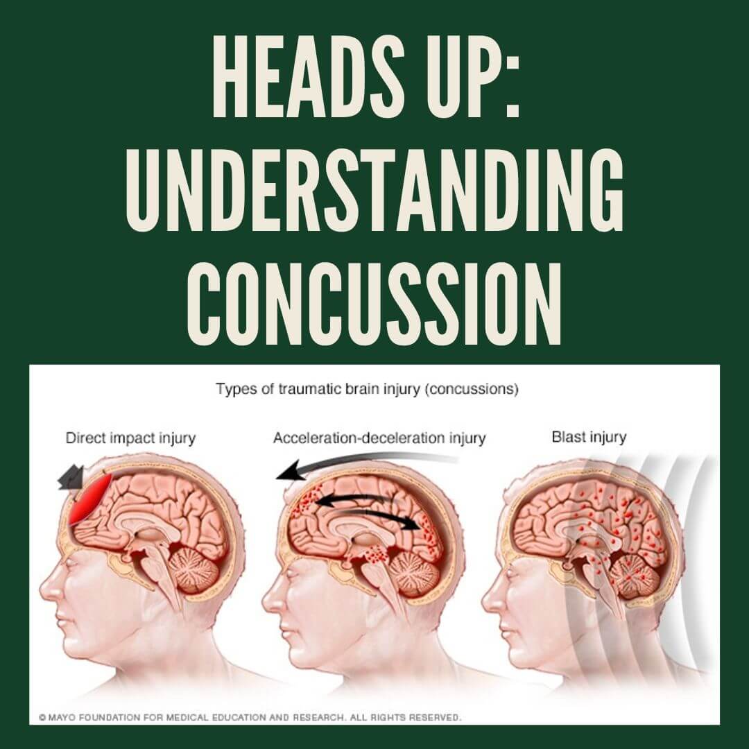
The May seminar for our Health Talks series, Practical Medicine: Empowering Women, Families, and Athletes, was “Active and Aware: How to manage acute sports injuries”. This was presented on 20th of May. In this presentation, Dr Tess was joined by podiatrist Ben Sweeting, and physiotherapist Alex Downie. They discussed common injuries of the ankle, knee and shoulder.
What do I do if I injury myself in sports?
If you sustain an injury while training or playing a sport, your first port of call may be your local emergency department, if your injury is severe enough. However, many injuries are milder and do not require paramedic/emergent care.
Many athletes will first call upon their local healthcare provider, such as their GP, physiotherapist, or podiatrist.
During the initial visit, your healthcare provider will ask about the event, including what happened in the current injury and any history of previous injuries. A physical examination will be followed by possible diagnoses, and you may need further investigations, including possible imaging (x-ray, ultrasound, CT scan, or MRI). Treatment may involve pain relief, taping or splinting, and referrals to sports physicians or orthopaedic surgeons. There will be a period of rehabilitation with a podiatrist or a physiotherapist before you can return to your usual level or training or play.
Injuries
Acute injuries
- Occur suddenly during a specific event
- Tend to have sharp pain, swelling, bruising
- Examples: joint sprain, muscle strain, ligament tears, fractures, dislocations, blisters
Chronic injuries
- Gradually happen over time due to overuse or misuse
- Have a dull ache, stiffness, swelling after activity
- Examples: tendinopathies, shin splints, stress fractures
All injuries go through four stages of healing
Stage 1: bleeding (0-24 hours) – starts immediately after an injury and sets the foundation for proper healing
Stage 2: Inflammation – Damaged cells release chemicals to stimulate inflammatory response; white blood cells remove damaged tissue. Symptoms: Swelling, redness, warmth, pain. Pain helps protect the area and prevents further damage
Stage 3: Proliferation (Day 2 to 3 Weeks) – New tissue starts to grow; cells build collagen and repair structures; new blood vessels form. Tissue is immature and not yet strong enough for full activity.
Stage 4: Remodeling (Weeks to Months) – Tissue matures and strengthens. Cells align fibers to handle stress and movement. This is the final stage for long-term recovery and function
Factors that Inhibit Healing
Age, genetic factors, poor nutrition, previous injury, metabolic and chronic diseases, e.g. diabetes and cardiovascular disease, obesity, activity levels (too little or too much), lifestyle factors like smoking, alcohol consumption, poor sleep etc.
Acute Injury Management
Most people are familiar with RICE – Rest, Ice, Compression and Elevation as the first steps to manage acute injuries.
This was superseded about 15 years ago by POLICE: Protection, Optimal Loading, Ice, Compression, Elevation.
Today, with modern recovery, show your acute injury PEACE & LOVE. This method covers acute, subacute, and chronic injury stage.
PEACE: Your Guide Right After Injury (Acute phase 0-2 weeks)
- Protect
- Elevate
- Avoid anti-inflammatories
- Compress
- Educate
Protect the Injury – But Don’t Rest Too Long. Limit movement for 1–3 days. This reduces bleeding and prevents further damage. But: avoid total rest — too much can weaken tissue
Elevate – It’s still worth it. Keep the injured limb above the heart. This helps reduce swelling.
Skip the Pills? Anti-inflammatories can slow long-term healing. Inflammation is a natural and necessary phase of healing.
Make sure to discuss pain and recovery with your GP – there are many instances in which pain relief and anti-inflammatories are indicated, especially if pain is impacting on your ability to complete your normal daily functions and activities.
Compression – Use it smartly. Taping or bandaging reduces bleeding and swelling, allows joint to keep full range of motion
Educate for Empowerment. Active recovery beats passive treatments. Set clear, realistic recovery goals. Avoid creating patient dependency
LOVE: Guiding Long-Term Recovery (> 2 weeks)
- Load
- Optimism
- Vascularization
- Exercise
Load: Use It – Don’t Lose It. Movement and exercise build tissue strength. Start loading as soon as pain allows. This will boost tissue remodeling.
Optimism: Healing Happens in the Brain Too. Mental health matters: fear, stress, and negative thinking delay recovery. Positive outlook = better outcomes.
Vascularization and Exercise: Get the Blood Flowing. Safe aerobic exercise boosts circulation, reduces pain, improves mood, enhances recovery without aggravating injury.
How to prevent acute sports injuries
🏃 Develop a balanced fitness plan: Include cardio, strength training & flexibility.
🧊 Warm up & cool down
🧘 Stretch regularly
👟 Use proper gear & supportive shoes for your sport.
📚 Have your biomechanics assessed and address any deficits
😴 Rest when tired.
🏋️♂️ Strength train with good form & full range of motion.
Lateral Ankle Sprains
Ankle Sprains – often involve damage to outer (lateral) ligaments of ankle. Mechanism often involves inversion of the ankle, jumping or landing, or even stepping on an opponents foot.
- Grade 1 = mild stretching
- Grade 2 = partial tear
- Grade 3 = full rupture
When do I need an X-ray? Ottawa Ankle Rules
You need an X-ray if:
You can’t put weight on your foot: If you can’t take 4 steps right after the injury
You have pain in specific bone areas: Pain is around the ankle (near the bump on either side)
- Tenderness on the bottom 6 cm of the outer ankle bone (fibula),
- Tenderness on the bottom 6 cm of the inner ankle bone (tibia).
Pain around the midfoot
- Tenderness over the base of the 5th toe bone (outer edge),
- Tenderness over the navicular bone (top inner side of the foot).
Rehabilitation aim to increase strength in the ankle, restore range and flexibility, and restore balance.
Knee Injuries
General Considerations
- Reporting a “pop”
- Inability to weight bear
- Inability to straighten the knee
- Inability to flex the knee past 90°
- Swelling immediately and the day after
Ottawa Knee Rules: indications for x-ray with acute knee injuries
- Age> 55
- Isolated patella tenderness
- Tenderness at head of fibula
- Inability to flex knee 90°
- Inability to weight bear (4 steps) immediately after injury and in emergency department
ACL Injuries
Usually associated with a “pop”. 85% in professional football (soccer) involve non-contact or indirect contact. There is a feeling of knee giving way, or “knee doesn’t feel right”. The athlete is not always completely incapacitated. Swelling will occur. If you are <50 years old and if there is sufficient suspicion, you can get bulk billed MRI scan via GP.
ACL Injury Management
- Surgery vs Conservative – a conversation to have with your orthopaedic surgeon
- Return to play times
- At least 9 months
- Ideally 12 months+ in adolescents
- Strict criteria to meet prior to return to sport
- Increased risk of 2nd ACL injury on same side and index injury on opposite side
Meniscus Injuries
After an injury to a meniscus, an athlete may feel the sensation of locking or catching, or an inability to straighten knee. This is often associated with other injuries or in isolation. You are eligible for a bulk billed MRI via your GP if you are < 50 years old with sufficient suspicion.
Meniscus Injury Management
- Surgery vs conservative
- Meniscal repair vs Meniscectomy (trim / removal)
- Good quality rehab required for return to sport
- Return to sport times
- 6-12 weeks meniscectomy
- 3-6 months meniscal repair
MCL Injuries
Typically involve a valgus force. Can be associated with ACL/Meniscus injuries. Isolated MCL almost always is managed conservatively. There is a 6 week period of bracing for grade 2-3 injuries. Return to play follows this period.
Other Injuries
- Patella Dislocation
- PCL injury
- Cartilage injury
Shoulder Injuries
AC Joint Injury
Involves a shoulder impact which causes acromioclavicular dislocation and the scapula to be pushed down.
Grades 1-5. Grade 2 or more has a visible deformity. There is tenderness to palpate. Athletes can report feeling a “pop”. There is a need to differentiate between ACJ and shoulder dislocation. X-ray is useful for grading and exclusion of fracture.
AC Joint Injury Management
- Period of immobilization in sling
- Surgery vs conservative
- Graded rehab
- High risk of recurrence / aggravation if returning early
- Can support with strapping / padding
Shoulder Dislocation
Similar mechanism to ACJ injury. Athletes will potentially feel a “pop”. There will be a visible deformity if the joint doesn’t spontaneously reduce. A suitably qualified physiotherapist / doctor may attempt relocation. If it doesn’t go easily then off to the emergency department. The instability may be subtle or subluxation vs dislocation (increasing stages of dislocation).
Shoulder Dislocation Management
- Sling immobilization for comfort. No difference in recurrence <1 week or > 3 weeks. (Lill et al,
2001) - Recurrence rates from 14-100%
- Male < 20yo = 86.6% at 5 years
- Risk increases if:
- Returning to contact or overhead sport
- Hypermobile
- Bony damage to shoulder
- Shoulder Stabilization
- Risk of recurrent instability after stabilization (bankart repair) 10-15%
- Risk of recurrent dislocation as low as 5% with Latarjet procedure (Bulleit et al, 2025)
- Immobilization often 6 weeks
- Average return to play time ~6 months if passing criteria
Health Talk Series
This blog is from our monthly in person events being run at Carina Leagues Club. Each month we focus on a different health topic.
For more information or to join us in person click here: